Speedy AI image analysis could help doctors during surgery
It compares 3D scans up to 1,000 times faster than before.

Right now, comparing 3D medical scans is a pain -- it can take two hours or more to see what's changed. And that spells trouble for surgeons, who may have to bring patients back to the operating room if a tumor removal wasn't a complete success. Thankfully, AI technology may eliminate that hassle. An MIT-led research team has crafted a machine learning algorithm that can analyze 3D scans up to 1,000 times faster than before, making it possible to study changes almost in real time -- less than a second on a PC with a fast graphics card.
The approach starts by training the algorithm on "thousands" of image pairs, teaching it how to align the scans and provide meaningful comparisons. After that, it can map every voxel (3D pixel) of both scans at the same time. That's no mean feat when there are frequently a million voxels between the two pictures. Existing systems start fresh with every new image, effectively forgetting everything they learned about location from the last time around.
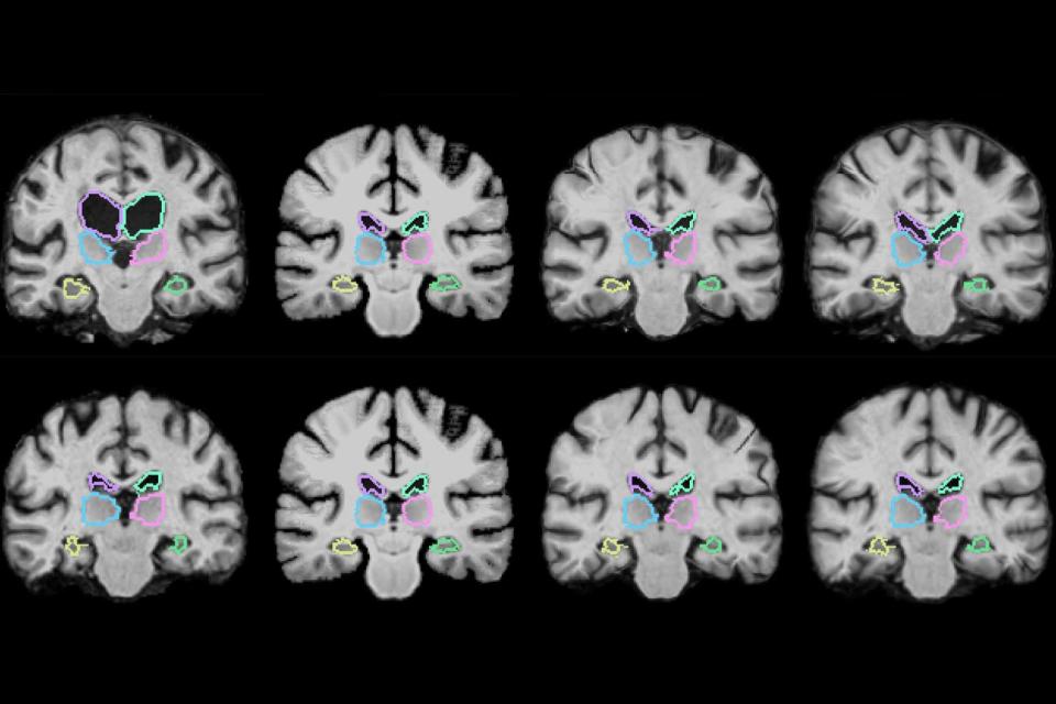
As you might have predicted, that speed-up would be extremely useful for surgeons, who could theoretically find out how successful a procedure was while they're still in the middle of surgery. It could be useful for much more, for that matter. While MIT has focused on brain scans, it could also be useful for lungs and any other organ where fast analysis could make life easier for both doctors and patients.
